Cherry Angioma Removal: Causes, Symptoms, Treatment & Recovery

A cherry angioma is a harmless cluster of tiny blood vessels located close to the skin's surface. These are not dangerous, and they are not a form of skin cancer. That's why most people don't need treatment at all.
Cherry Angioma Image
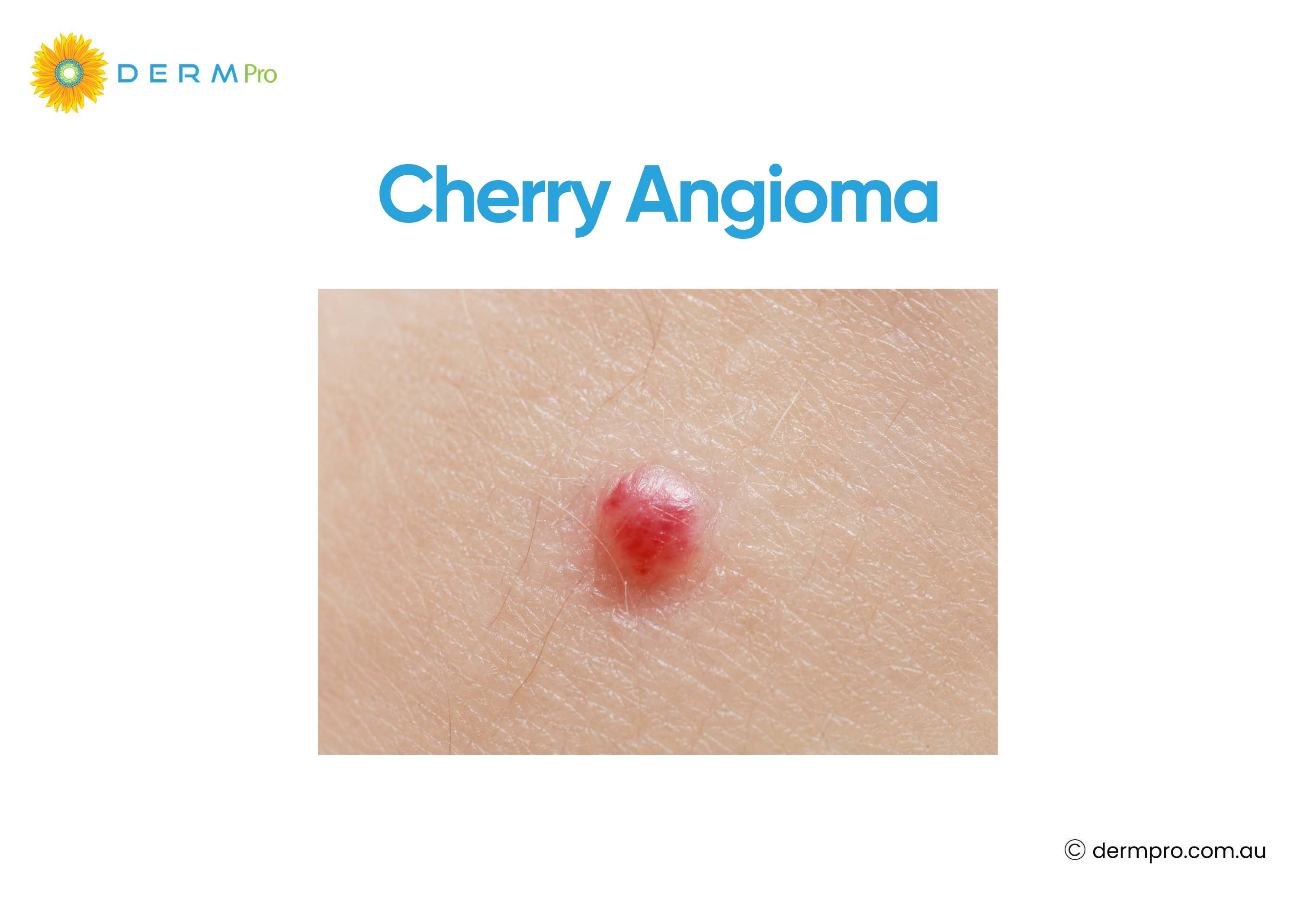
If yours is not bothering you, you can simply leave it alone. But if it catches on clothing, bleeds, or you just don't like how it looks, a doctor can remove it quickly and safely.
The most effective removal options are laser therapy, electrocautery, cryotherapy, and shave excision. Each works well, and your doctor will choose the best one based on the size, colour, and location of the spot.
See a doctor immediately if a spot changes shape, grows quickly, has an uneven border, or looks different from your other angiomas. These signs don't automatically mean something serious, but they are worth checking.
What Is a Cherry Angioma?
Angiomas are small, benign growths made up of blood vessels. A cherry angioma, sometimes called a cherry hemangioma, is the most common type. It forms when tiny blood vessels bunch together close to the surface of the skin.
Cherry Angiomas
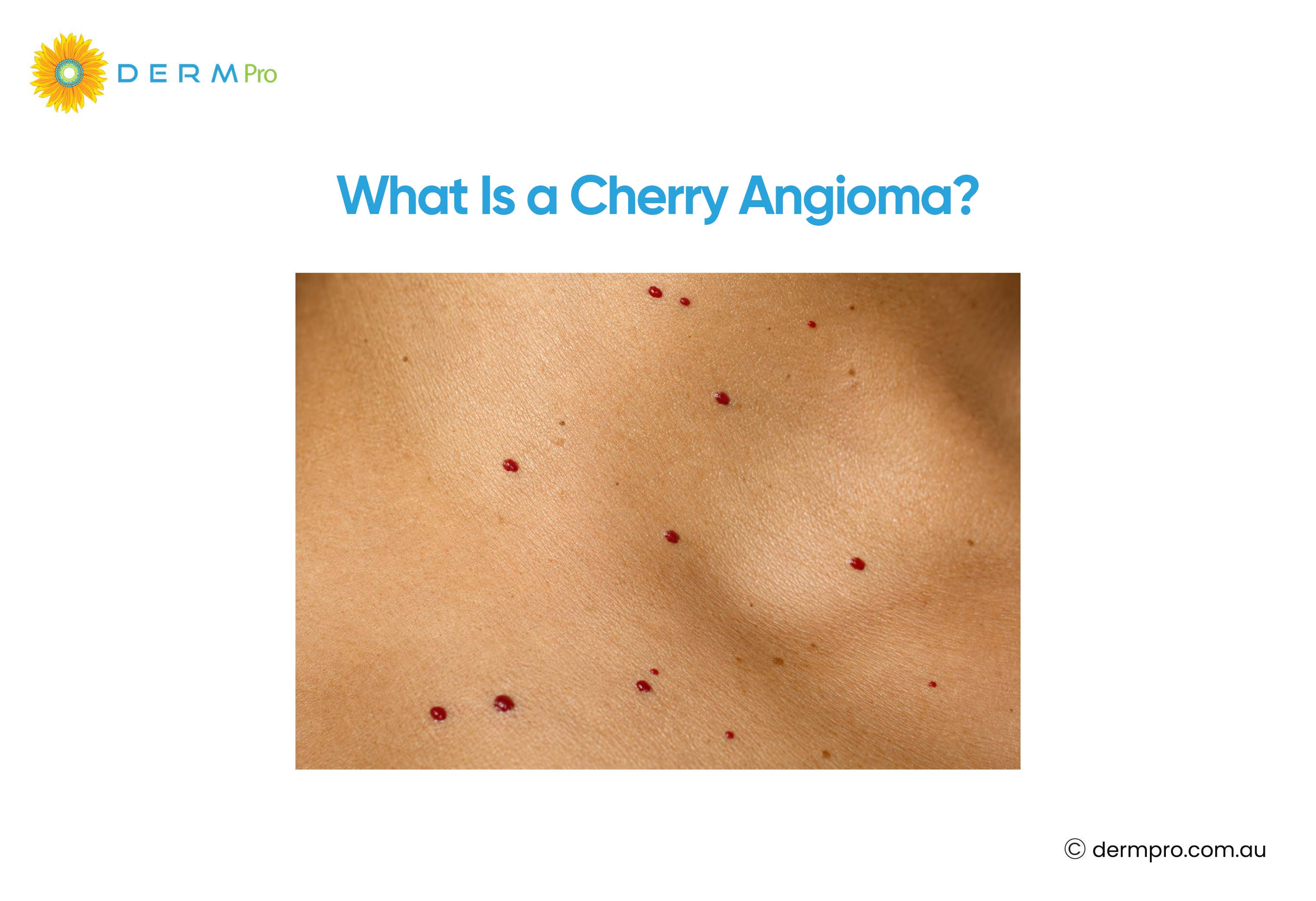
People often call them red moles or red freckles because of their round shape and bright colour. Despite the name, they are not actually moles, since moles are made of pigment cells rather than blood vessels. However, they are also known as Campbell de Morgan spots, cherry hemangioma or Senile Angiomas.
An angioma of the skin usually looks like a small, smooth, dome-shaped or flat red dot. It can range from the size of a pinhead to around a centimetre across. Colour can vary from bright cherry red to a deeper purple-red.
Cherry angiomas can appear anywhere on the body, but they most often appear on the chest, back, arms, and abdomen. It is common to have several at once, and new ones often appear as people get older.
Cherry Angioma Removal Options
Doctors usually remove cherry angiomas with laser treatment, electrocautery, cryotherapy, or shave excision. The best treatment depends on the size, colour, location, and number of angiomas you have.
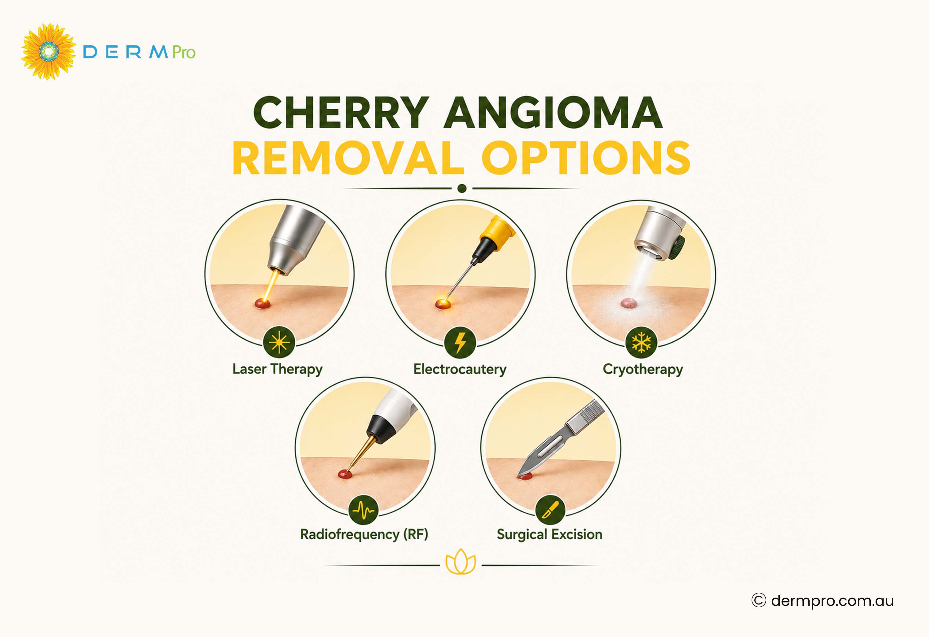
Laser therapy
This method uses a focused beam of light to target the blood vessels inside the angioma. The heat from the laser seals the vessels, and the spot fades over the following weeks. This method works well for small to medium angiomas and usually leaves minimal marking.
Electrocautery
This treatment uses a fine heated probe to burn away the angioma. It works quickly and controls bleeding at the same time. Your skin may need a week or two to heal fully.
Cryotherapy
Cryotherapy is a process that freezes the angioma with liquid nitrogen. The tissue dies and flakes away over the following days. It is a quick in-office treatment. This option suits smaller, flatter angiomas best.
Shave excision
This method involves shaving the angioma off with a small blade under local anaesthetic. This method suits larger or raised angiomas and lets your doctor send a sample for testing if needed.
|
Procedure |
How it works |
Advantages |
Recovery Time |
Possible side effects |
|
Laser Therapy |
Light energy seals the blood vessels |
Precise, minimal scarring |
A few days to two weeks |
Mild redness, temporary darkening |
|
Electrocautery |
Heat destroys the tissue |
Fast, controls bleeding well |
One to two weeks |
Small scab, slight scarring risk |
|
Cryotherapy |
Freezing destroys the tissue |
Simple, no cutting involved |
One to two weeks |
Blistering, temporary skin lightening |
|
Shave Excision |
Blade removes the raised tissues |
Allows tissue sampling |
One to three weeks |
Small scar, mild bleeding |
Which treatment works best for removal of cherry angioma?
Laser therapy tends to give the most consistent cosmetic results for small angiomas. However, larger or raised angiomas often respond better to shave excision.
Which treatment leaves the least scarring to remove angiomas?
Vascular Laser Therapy, including pulsed dye lasers such as V-Beam and KTP lasers, leaves the least scarring to remove angiomas. Because they can target the haemoglobin within the abnormal blood vessels while limiting damage to surrounding skin. Cryotherapy is another treatment option in this aspect, but the risk of visible marks or pigment changes can vary depending on the treatment technique and individual skin type.
Which treatment works best for larger angiomas?
Shave excision or electrocautery usually handle larger or raised angiomas more effectively than laser alone.
How long does recovery take?
Most people heal within one to two weeks. However, full fading of any redness can take a little longer.
How to Remove Cherry Angiomas at Home (Is it Safe or Not?)
Many people search for ways to remove cherry angiomas at home. This makes sense. A quick fix sounds appealing, and some people feel embarrassed booking an appointment for something so small.
Dermatologists strongly discourage home removal. Cherry angiomas are full of blood vessels, so cutting, picking, or using acid-based products can cause heavy bleeding. Skin near an angioma is also delicate, so home treatments raise the risk of infection and lasting scars.
There is another reason to avoid self-treatment. A doctor can confirm the spot is actually a cherry angioma and not something else that needs closer attention. Removing a spot at home skips this important diagnostic step.
If you want a red spot removed, booking an appointment with your GP or a dermatologist is important. The procedure takes only minutes and carries far less risk.
What are the Causes of Cherry Angiomas
Researchers haven't pinned down one single cause of cherry angiomas. Several factors appear to play a role.
- Ageing. Cherry angiomas become more common after age 30 and tend to increase in number with age.
- Genetics. Cherry angiomas appear more often in some families, which suggests that inherited genes may play a role.
- Hormonal changes. Shifts in hormone levels can trigger new angiomas or make existing ones more noticeable.
- Pregnancy. Hormonal changes during pregnancy can lead to new angiomas appearing.
- Environmental factors. Some researchers have looked at climate and chemical exposure as possible contributors, though evidence remains limited.
- Ongoing research. Scientists continue to study why blood vessels cluster this way and why some people develop many more angiomas than others.
Cherry Angioma Symptoms
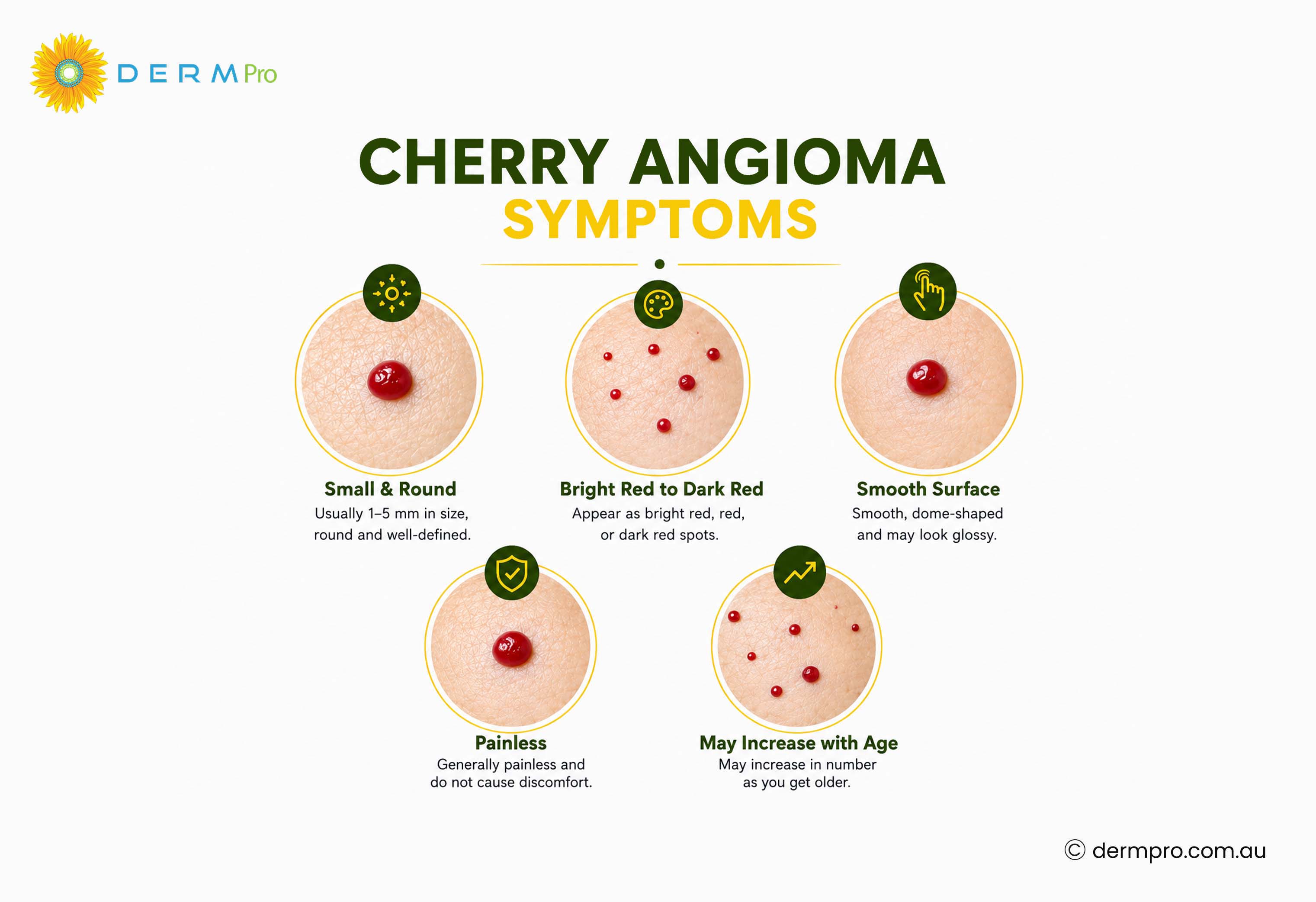
Cherry angioma red spots on skin usually look smooth, round, and bright red. They rarely cause pain or itching.
Red freckles on skin are simply another common name people use for the same spots. They are flat or slightly raised and don't spread like a rash.
Cherry angioma bleeding can happen if the spot is scratched, caught on clothing, or knocked. Because it is made of blood vessels, it can bleed more than you might expect from such a small mark. Firm pressure with a clean cloth usually stops it.
Cherry angioma turning black can happen after an injury, when trapped blood clots inside the angioma. This is usually harmless, but any sudden colour change is worth showing to your doctor to rule out other causes.
Cherry angioma on the scalp can be harder to spot under hair and may bleed more when disturbed by brushing or washing.
Cherry angiomas on the legs are common and can sometimes be mistaken for bruises or other vascular marks.
Flat angiomas stay level with the surrounding skin rather than forming a raised bump. They still keep the same bright red colour as raised angiomas.
Cherry Angioma vs Melanoma
Cherry angiomas and melanoma look very different once you know what to check. Melanoma is a form of skin cancer, and it can vary a great deal from person to person, so always have any unusual spot checked by a doctor rather than relying on comparison alone.
|
Feature |
Cherry Angioma |
Melanoma |
|
Colour |
Bright red, purple red, smooth and well-defined |
Often brown, black, or multiple colours |
|
Size |
Usually under 1cm |
Often larger than 6mm and growing |
|
Shape |
Round, symmetrical |
Often irregular or asymmetrical |
|
Border |
Smooth, well-defined |
Uneven, blurred, or notched |
|
Growth Pattern |
Slow, stable over time |
Can grow or change quickly |
|
Bleeding |
Only after injury |
Can bleed without injury |
|
Cancer Risk |
Not cancerous |
Cancerous, needs urgent care |
|
Medical Evaluation |
Optional, for comfort or appearance |
Essential and urgent |
If a spot shows any of the melanoma features above, or changes in size, shape, or colour.
Book a skin check with DermPro as soon as you can.
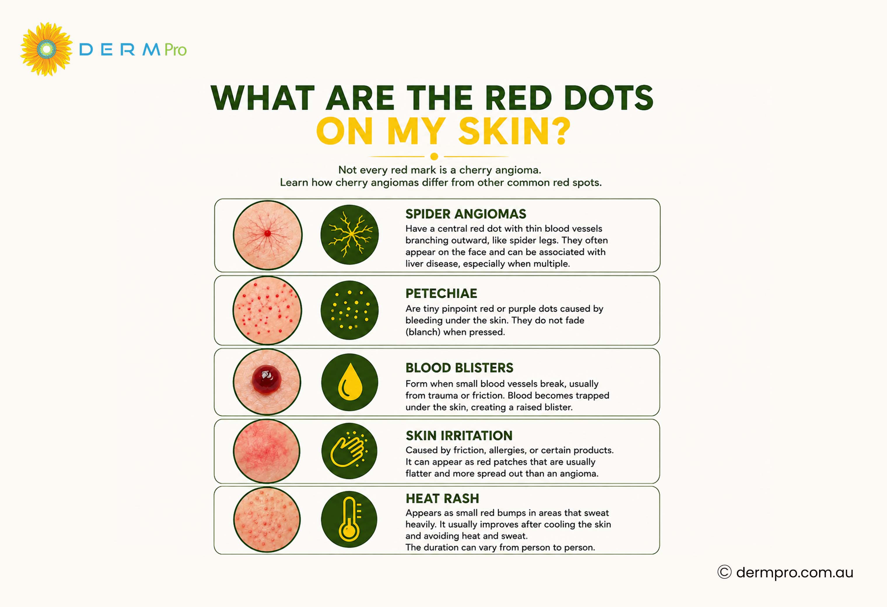
What Are the Red Dots on My Skin?
Not every red mark is a cherry angioma. Learn how cherry angiomas differ from other common red spots.
- Spider angiomas have a central red dot with thin blood vessels branching outward, like spider legs. They often appear on the face and can be linked to liver conditions.
- Petechiae are tiny pinpoint red or purple dots caused by bleeding under the skin. They don't fade when pressed, unlike many other red marks.
- Blood blisters form after an injury and contain trapped blood under a raised bubble of skin.
- Skin irritation from friction, allergies, or products can cause red patches that are usually flatter and more spread out than an angioma.
- Heat rash appears as small red bumps in areas that sweat heavily and usually clears within a few days.
How Doctors Diagnose Cherry Angiomas?
Doctors usually diagnose cherry angiomas quickly.
- Visual examination. Most cherry angiomas are recognisable at a glance because of their colour, shape, and texture.
- Dermoscopy. Your doctor may use a handheld magnifying tool with light to look more closely at the blood vessel pattern.
- Skin biopsy. In rare cases, if a spot looks unusual, your doctor may take a small tissue sample to confirm the diagnosis and rule out other conditions.
Recovery After Cherry Angioma Removal
Recovery from cherry angioma removal is usually straightforward.
- Recovery time. Most people notice healing within one to two weeks, depending on the method used.
- Skin care instructions. Keep the area clean and dry, and follow any dressing instructions your doctor gives you.
- Activities to avoid. Avoid picking at the area, and steer clear of swimming pools or heavy sweating until the skin has healed.
- Signs of infection. Watch for increasing redness, warmth, swelling, or pus, and contact your doctor if these appear.
- Follow-up care. Attend any follow-up appointment your doctor recommends, especially if a tissue sample was taken for testing.
Frequently Asked Questions
What are angiomas?
Angiomas are benign growths made of blood vessels. Cherry angiomas are the most common type found on the skin.
Are cherry angiomas cancerous?
No. Cherry angiomas are not cancerous and don't turn into skin cancer.
Can cherry angiomas disappear naturally?
Cherry angiomas rarely disappear on their own. Most stay the same size or grow slowly over time.
Can stress cause cherry angiomas?
There is no strong evidence that stress directly causes cherry angiomas. Ageing, genetics, and hormones play a bigger role.
Can children develop cherry angiomas?
Cherry angiomas are uncommon in children and appear far more often in adults over 30.
Can cherry angiomas return after treatment?
The treated angioma itself rarely returns, but new angiomas can appear elsewhere on the skin over time.
Are angioma pictures useful for diagnosis?
Pictures can help you compare general appearance, but they cannot replace an in-person diagnosis from a doctor.
What should I do if a cherry angioma starts bleeding?
Apply firm, gentle pressure with a clean cloth until the bleeding stops. If it doesn't stop within a few minutes, or it bleeds often, see your doctor.
Monitor Your Skin With Confidence with DermPro
Download the DermPro app to track new and changing spots over time, complete regular skin checks, and stay proactive about your skin health. If you notice anything unusual, seek advice from a qualified healthcare professional as soon as possible.
Conclusion
Cherry angiomas are a common and harmless part of skin ageing. They form when small blood vessels cluster near the surface of the skin, and most people develop at least one over their lifetime. Cherry angiomas are benign growths and are not considered cancerous. However, any spot that changes rapidly in appearance should be assessed by a healthcare professional.
If your cherry angioma doesn't bother you, there is no medical need to remove it. If you would prefer it gone, laser therapy, electrocautery, cryotherapy, and shave excision all offer safe and effective removal with minimal downtime. Avoid removing angiomas at home, since this raises the risk of bleeding, infection, and scarring.
The most important habit is simply paying attention to your skin. Get to know what your angiomas look like normally, and see a doctor promptly if you notice any changes in size, shape, colour, or texture.
This article is intended for informational purposes only and should not be considered medical advice, diagnosis, or treatment. Always consult a qualified healthcare professional regarding any concerns about your skin.
References
- American Academy of Dermatology: Information on cherry angiomas and skin health.
- Cleveland Clinic: Clinical guidance on cherry angiomas and treatment options.
- Mayo Clinic: Information on skin lesions, diagnosis, and treatment.
- Cancer Council Australia: Guidance on recognising suspicious skin changes.
Was this article helpful?
Your feedback helps us create better content for everyone.