Types of Moles and Skin Lesions: Signs & Differences

Almost every one of us has moles on our skin. Moles and skin lesions are so common that the average adult has between 10 and 40 of them. They can appear almost anywhere on the body, from the scalp to the soles of the feet.
It is important to know the types of moles on the skin and understand what to look for in skin changes.
Most moles do not cause problems and are harmless. However, some changes in moles can be worth discussing with a doctor. Early awareness is crucial to keep your skin healthy nowadays.
This blog explains the different types of moles, how to tell them apart from other skin lesions, and what signs are worth keeping an eye on. It also covers how tools like DermPro can help you monitor your skin over time.
What Is a Mole?
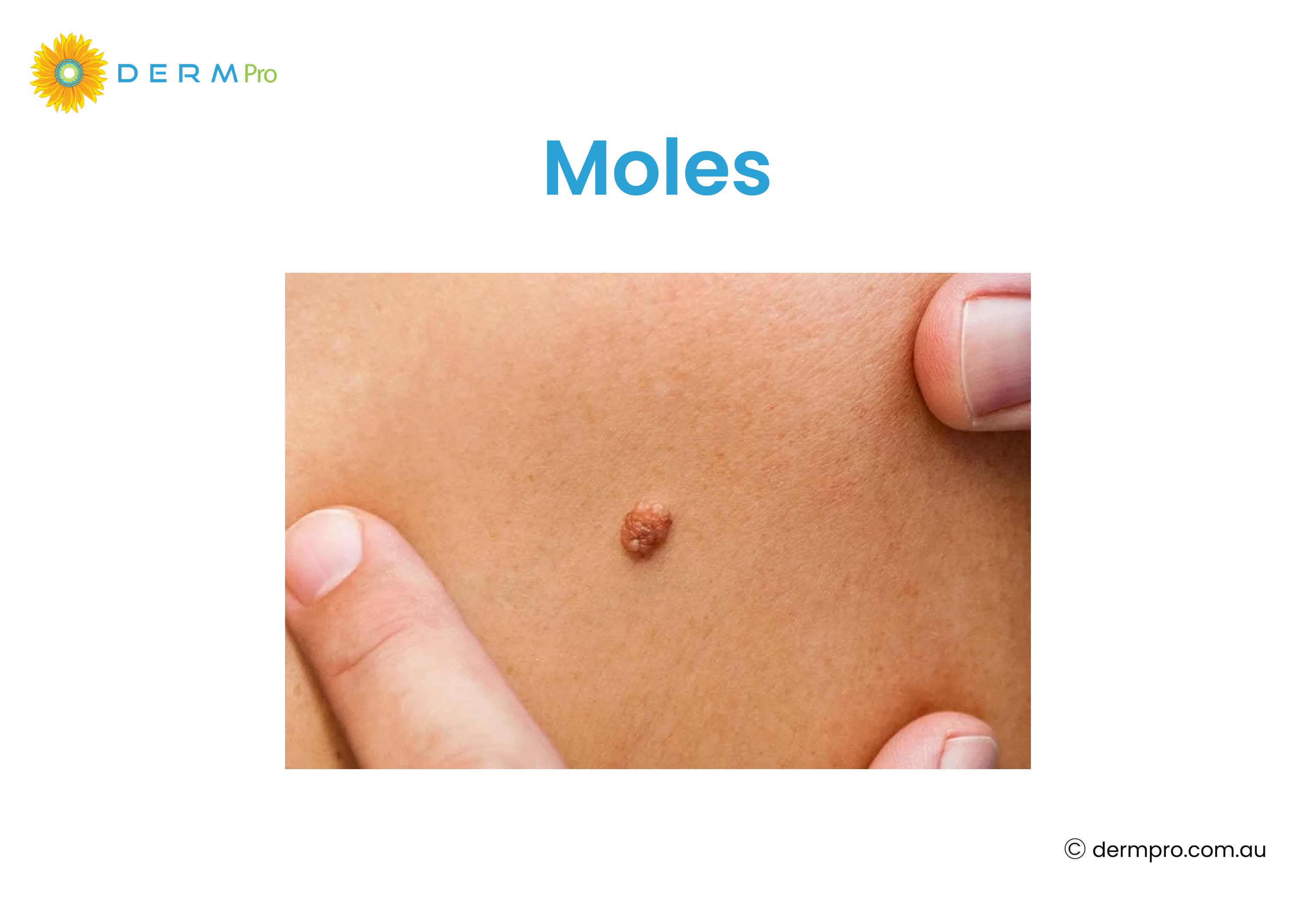
A mole is a growth on the skin that forms when melanocytes (pigment-producing cells) cluster together rather than spreading out evenly. Most moles appear during the first 30 years of life. Some people are born with them, while others may develop as they grow older.
Moles on the skin are usually round or oval. They are smaller than 6 millimetres (¼ inch) in diameter and have a smooth and consistent colour. They can be flat, raised, smooth or wrinkled. The medical term for a mole is a melanocytic naevus. They can be black, blue, pink, tan and brown in colour. Moles can appear anywhere on your body, including under the nail, scalp, toe, armpits and between your toes and fingers.
Some factors, such as sun exposure, genetics, and hormonal changes (such as during puberty or pregnancy), can all influence how many moles a person develops or how existing ones change.
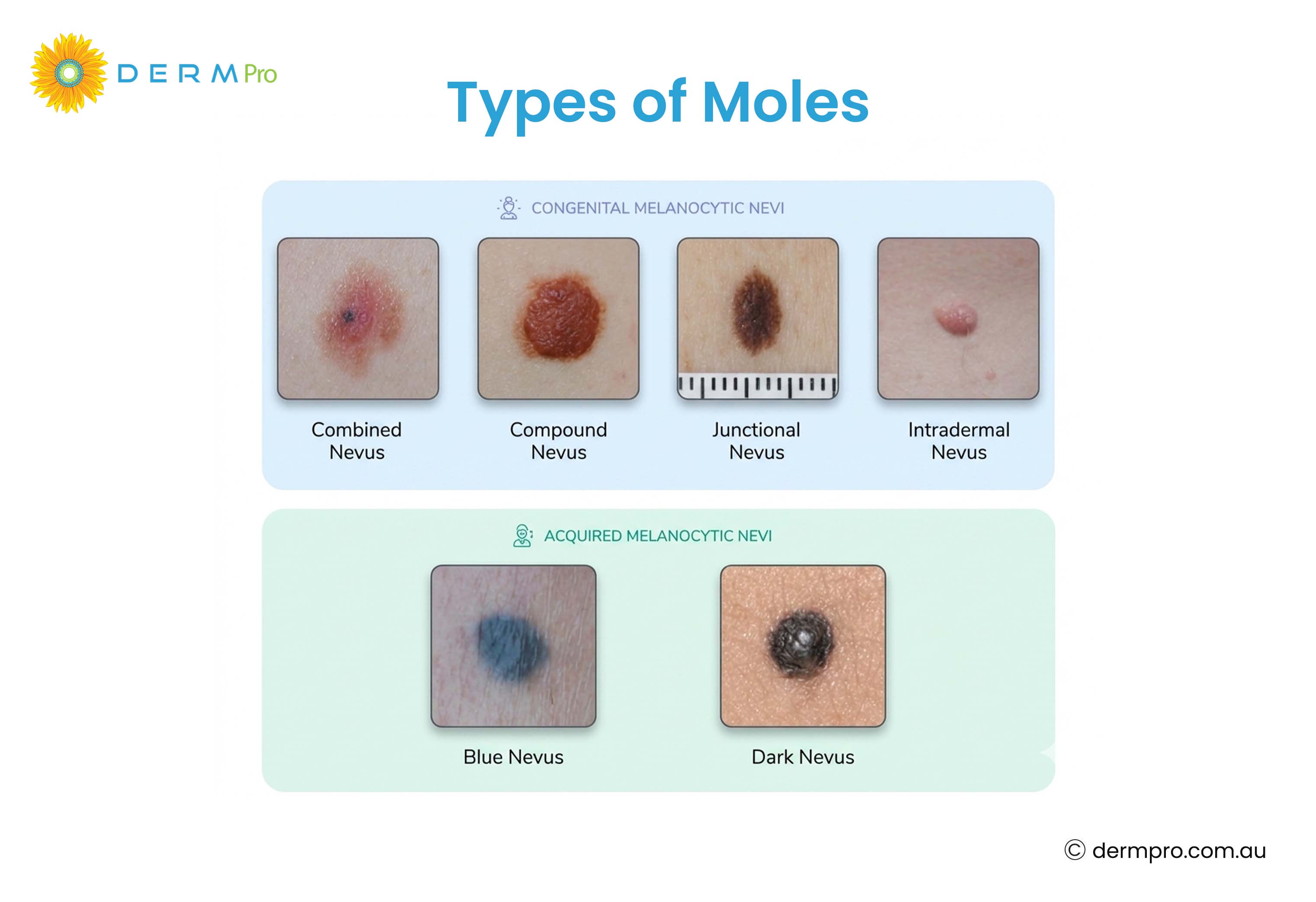
How Many Different Types of Moles Are There?
There are quite a few different types of moles. Understanding the differences helps you know what is normal and what might need attention. Broadly, moles fall into two categories:
- Benign (non-cancerous)
- Atypical (unusual or dysplastic).
Within these categories, there are many distinct subtypes.
Most moles are benign. Atypical moles look different from ordinary moles and may require closer monitoring or removal. But having atypical moles does not mean a person has or will develop cancer.
Types of Moles on Skin
Here are the main types of moles and what makes each one distinct.
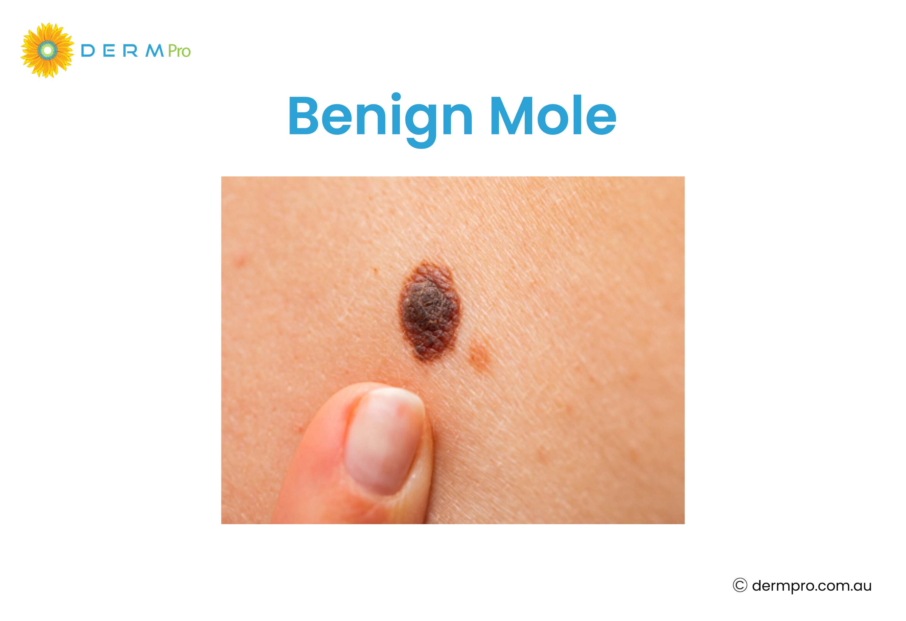
Benign Mole (Melanocytic Naevus)
A benign mole is a non-cancerous growth that poses no risk to health. It is typically uniform in colour (ranging from pink to brown or black). A benign mole has a well-defined border that stays the same over time. Most moles that people have fall into this category. The term "benign cancer mole" is sometimes used in searches, but it is worth clarifying that a benign mole is not cancerous at all and it is medically incorrect.
Benign Mole Image
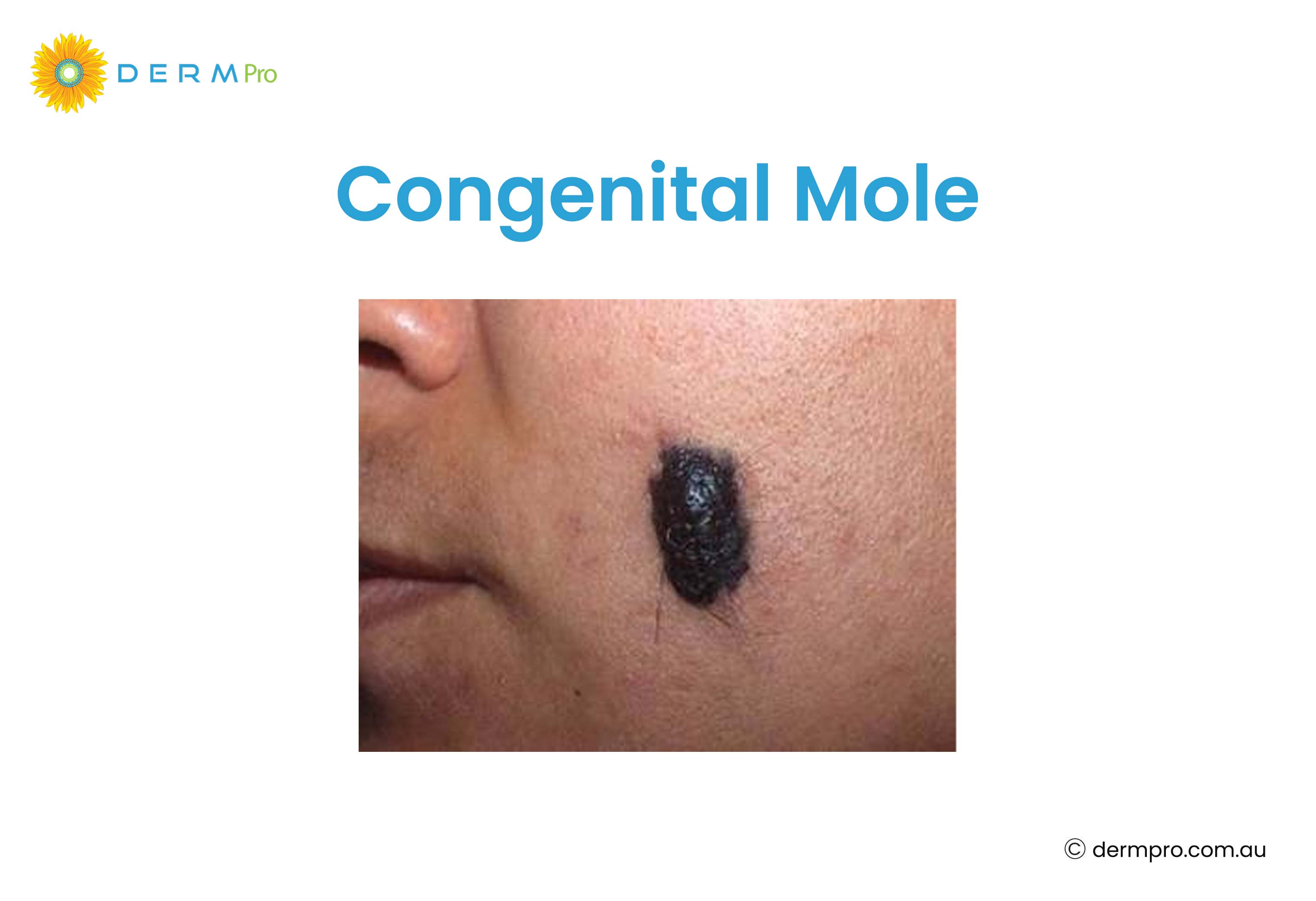
Congenital Mole (Congenital Melanocytic Naevus)
A congenital mole is a mole that is present at birth. It is also called a congenital naevus. It forms due to the proliferation of melanocytes. These occur in about 1 in 100 newborns. They can range from small marks to larger patches and sometimes have a slightly different texture. These naevi can vary in colour, size, and texture and may be flat or raised.
Larger congenital naevi (over 20 cm in adults) have a higher, though still small, risk of developing into melanoma. That's why regular monitoring of the skin is important to detect early changes suggestive of melanoma.
Congenital Mole Picture
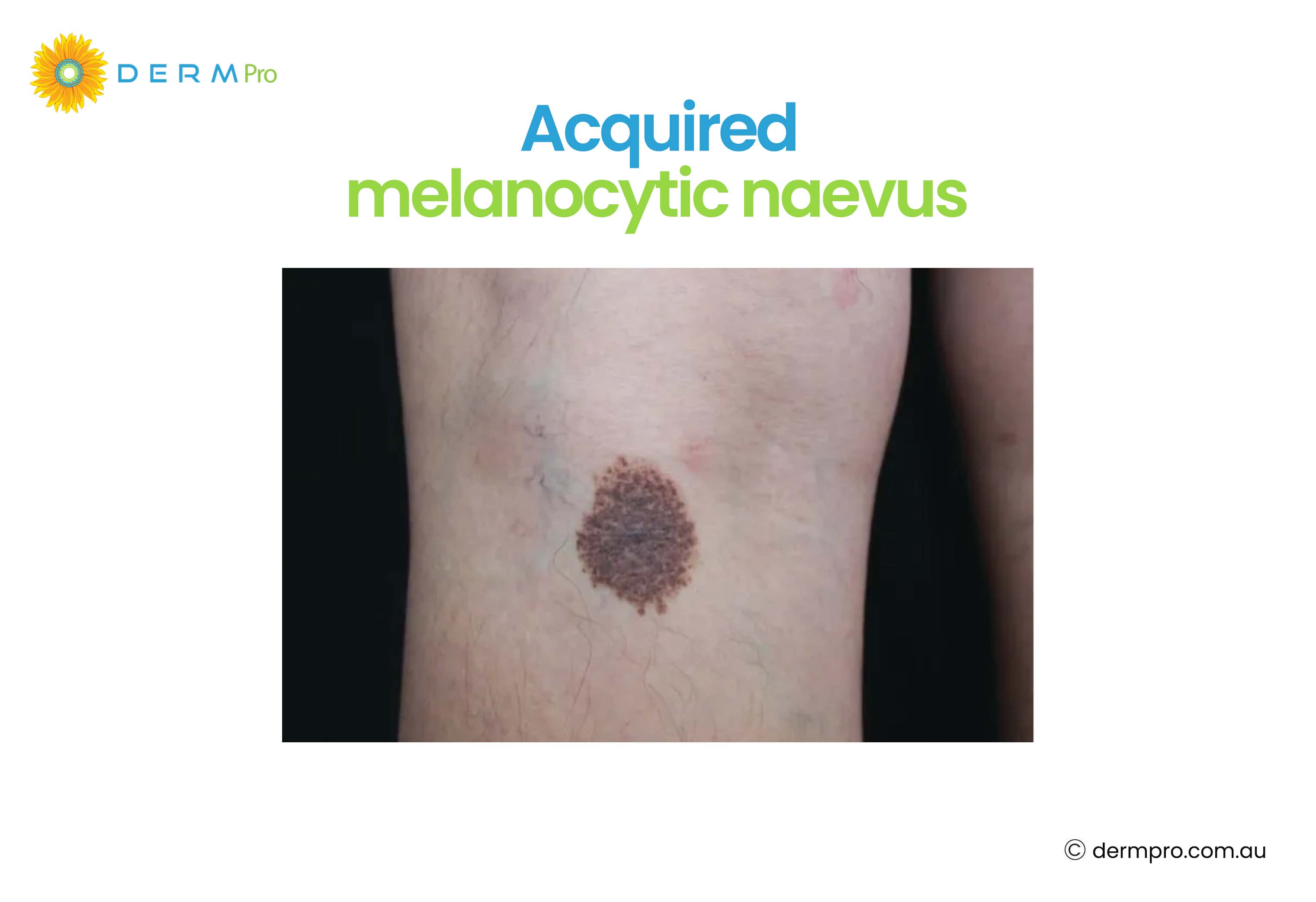
Acquired Melanocytic Naevus
An acquired melanocytic naevus is a mole that develops after birth. This is the most common type of mole. These moles develop in response to sun exposure, hormonal changes, and genetic factors. They typically appear during childhood or early adulthood and are almost always benign.
Acquired melanocytic naevus image
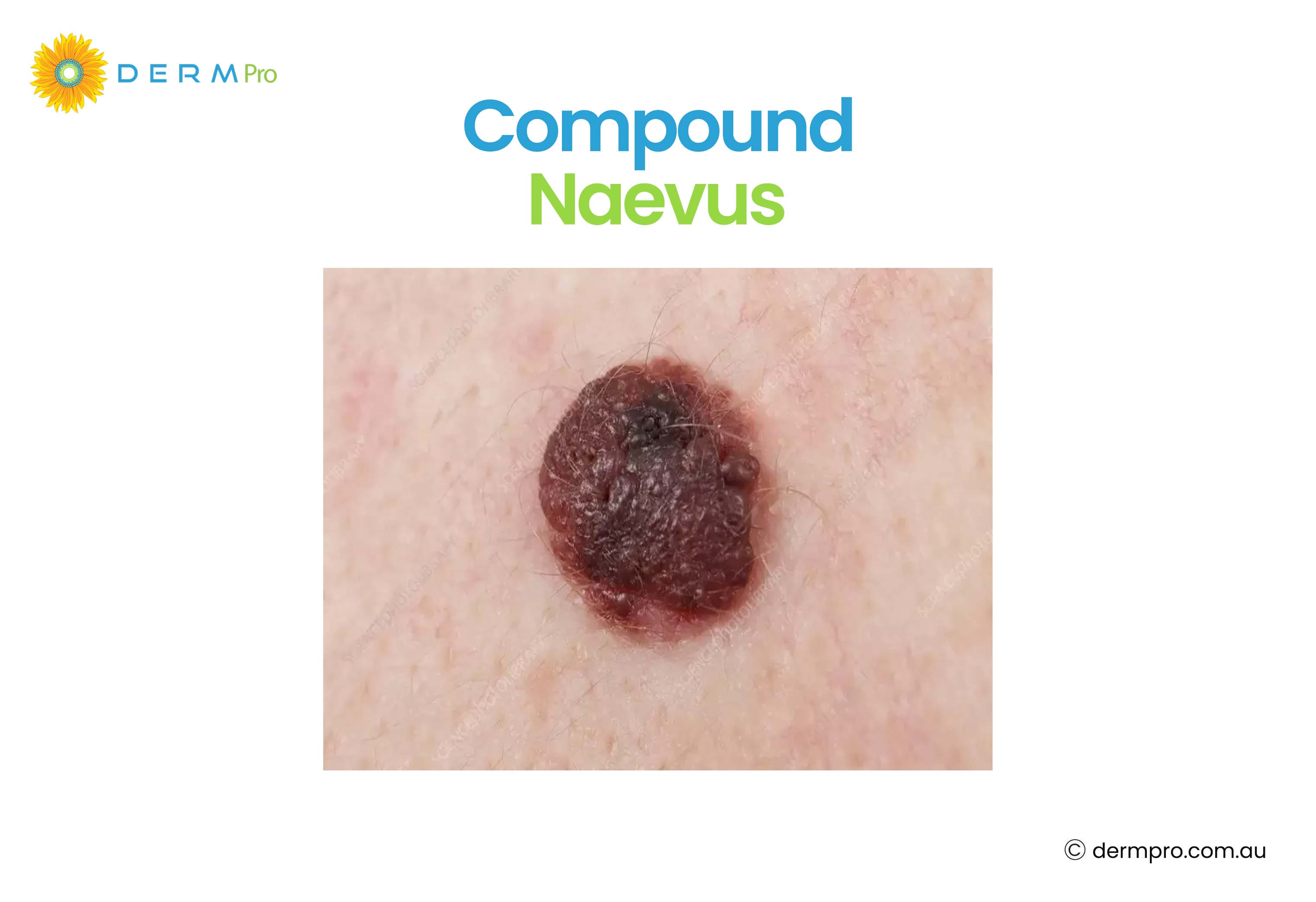
Compound Naevus
Compound naevi are acquired nevi. They contain melanocytes both at the junction between the epidermis and dermis and within the dermis itself. These moles are often raised and range in colour from tan to dark brown. They are generally benign.
Compound Naevus Image
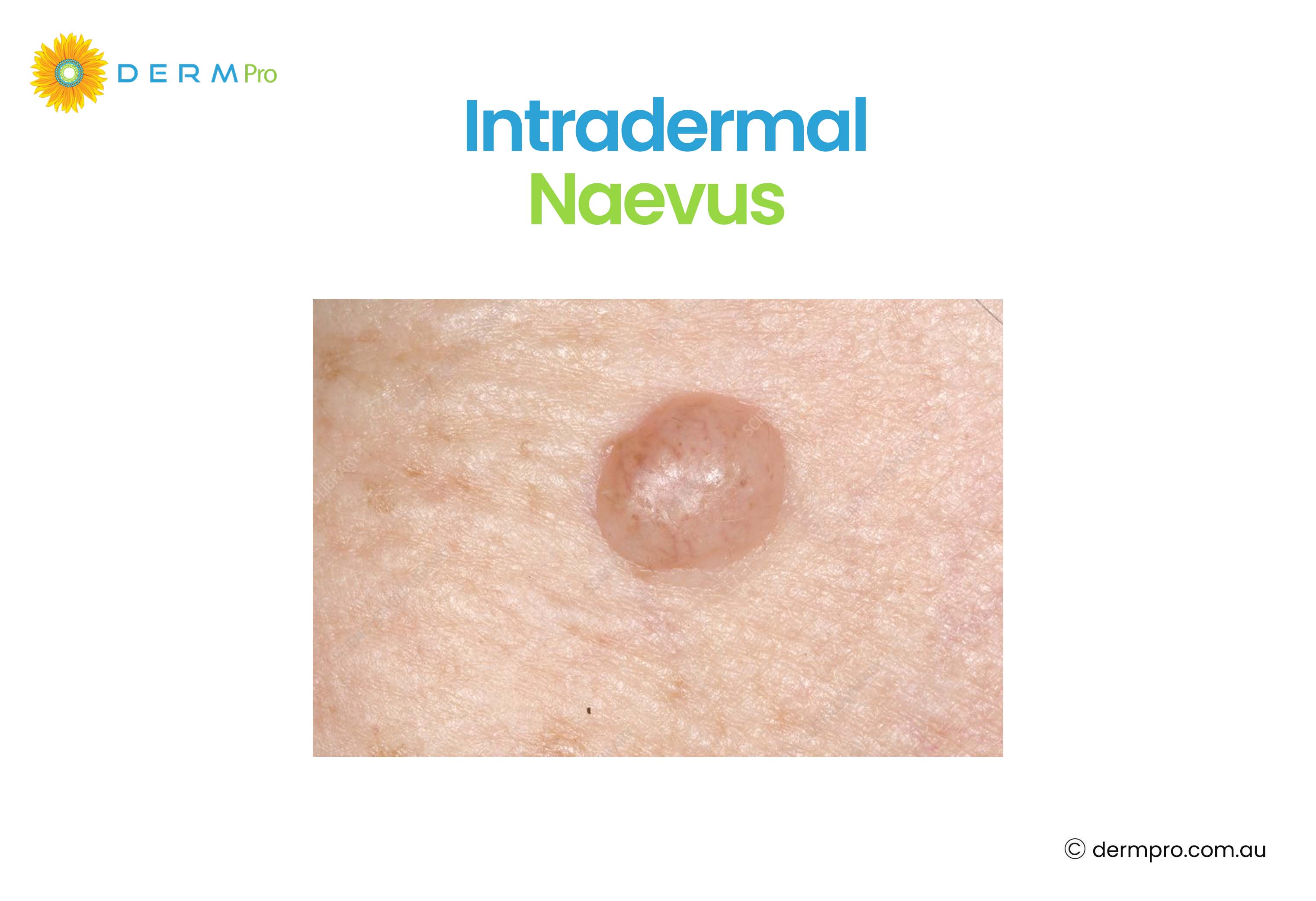
Intradermal Naevus
An intradermal naevus is an intradermal melanocytic naevus that sits entirely within the dermis. These are usually flesh-coloured or light brown. Intradermal nodules are often dome-shaped and are more common in adults. They tend to be soft to the touch. Because they sit deeper in the skin, they often appear less pigmented than other moles. Dermal nevi is another name for intradermal nevi.
Intradermal Naevus Picture
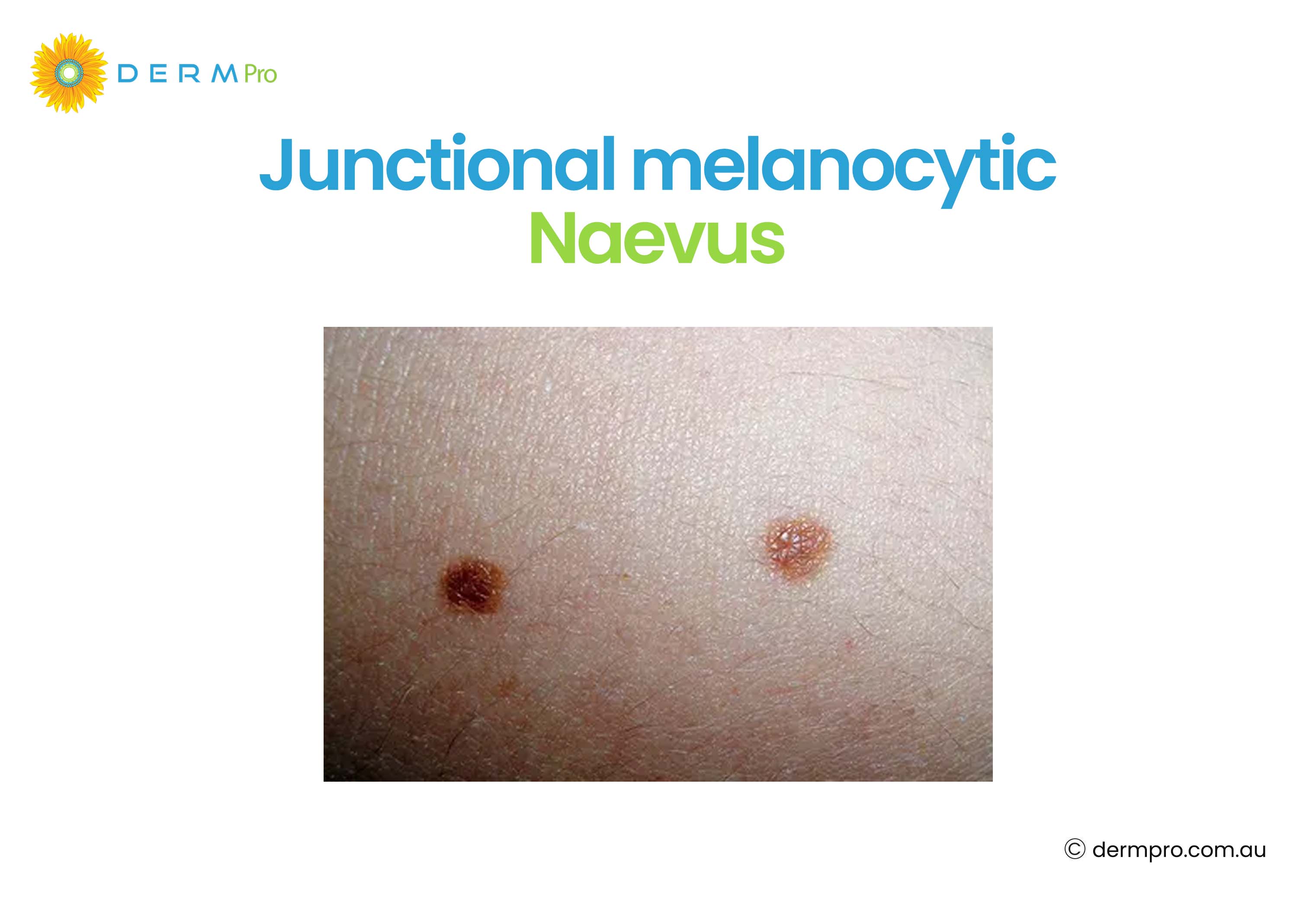
Junctional Melanocytic Naevus
A junctional melanocytic naevus develops where the epidermis and dermis meet. These are usually flat, dark brown or black, and have a smooth surface. They are common in children and young adults. Most remain stable and benign throughout life. Also, they are commonly found on arms, nails, feet, soles, legs, trunk and genitals.
Junctional melanocytic naevus image
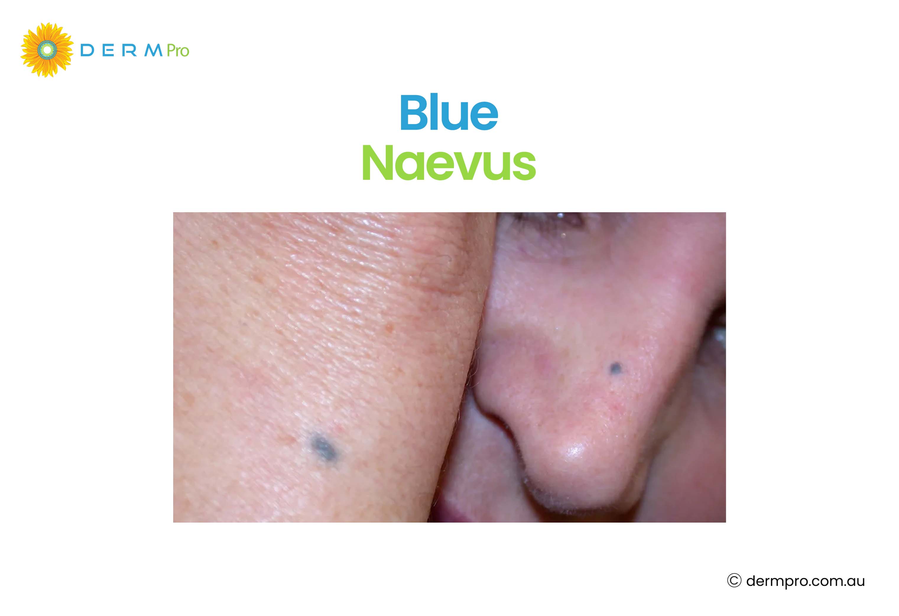
Blue Naevus Mole
A blue naevus mole gets its distinctive blue or blue-grey colour from melanocytes located deep within the dermis. The depth of the pigment causes the bluish appearance due to the way light interacts with the skin. Commonly they find in the neck, back of hands, feet, head and sacral area. But they can be in other parts of the body too.
Blue naevi are almost always benign and usually remain stable over time. Some studies suggest blue naevi may occur more commonly in women and in certain ethnic populations.
Common blue naevus or cellular blue naevus are categories of blue naevus mole. Common blue naevus has a smooth surface with a dome or flat shape. Its size ranges from 0.5 to 1cm. Cellular blue naevi are often larger than common blue naevi and may exceed 1 cm.
Blue Naevus Mole
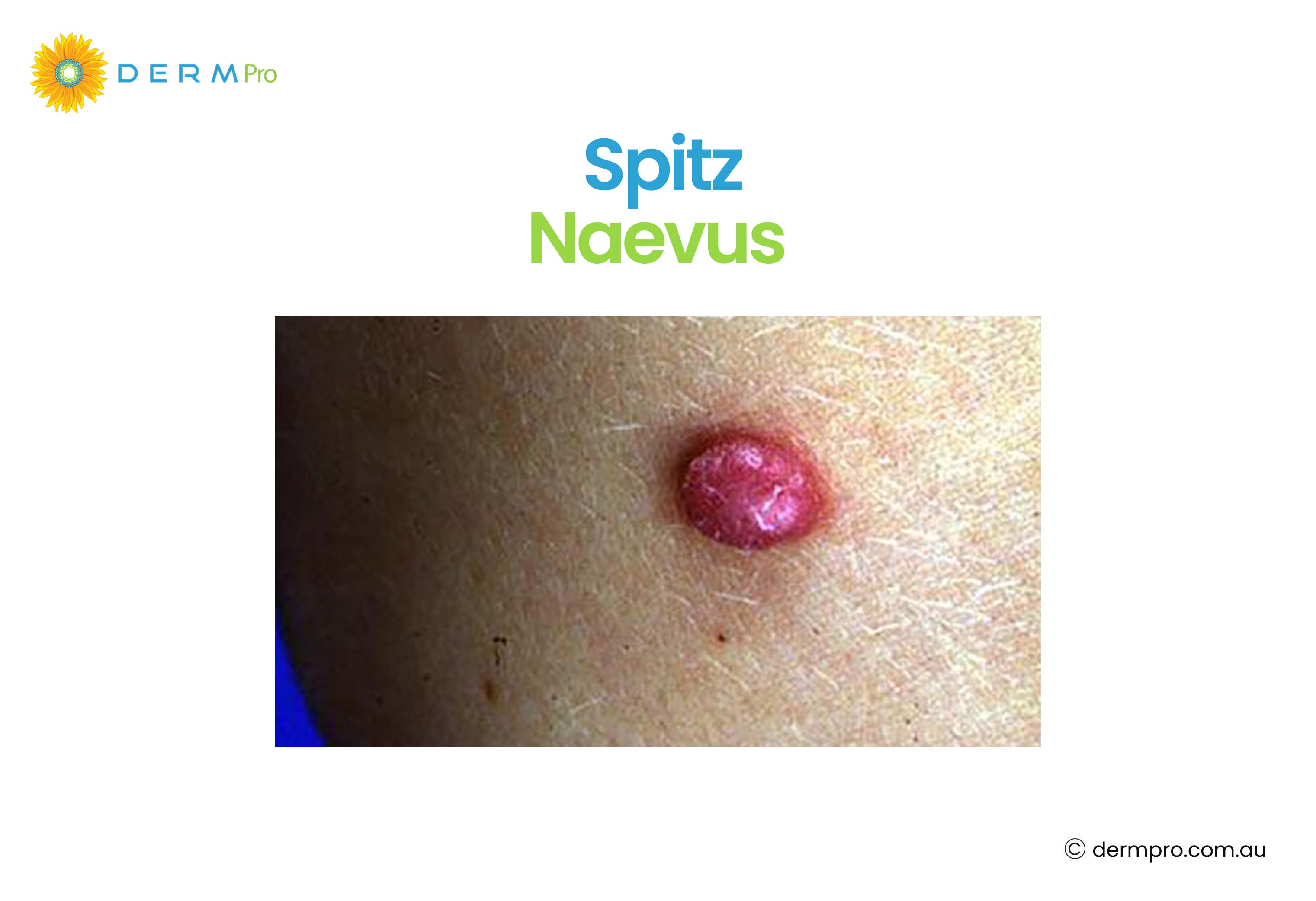
Spitz Naevus
A Spitz naevus is a type of mole. It commonly appears in children and teenagers. It is often pink, red, or brown, raised, and can grow quickly, which sometimes raises concern. However, most Spitz naevi are benign. Usually, they appear on the neck, leg, face and also on the shoulder, trunk and arm. Within 6 months, a spitz naevus can be 1 cm in size as they grow rapidly. A dermatologist will often recommend removal and biopsy to confirm the diagnosis, since they can look similar to melanoma under the microscope.
Spitz naevus image
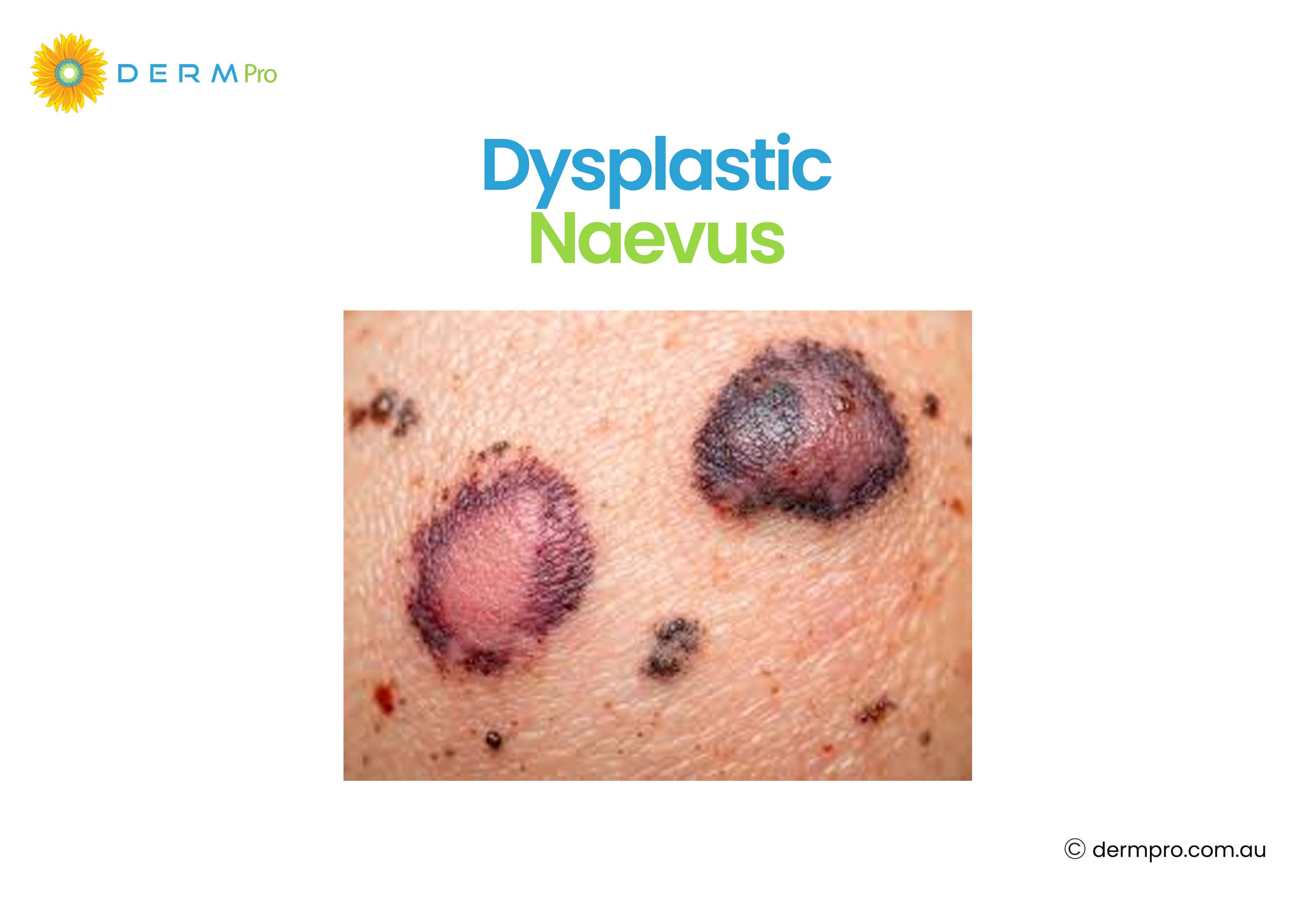
Dysplastic Naevus (Atypical Mole)
A dysplastic naevus atypical mole, also called an atypical naevus. It looks different from ordinary moles. These moles are often larger, have irregular or blurry borders, and contain multiple shades of colour. They do not always become cancerous, but people with multiple dysplastic naevi have a higher risk of developing melanoma over their lifetime.
Dysplastic Naevus Image
Compound Dysplastic Naevus With Mild Atypia
A compound dysplastic naevus with mild atypia shows low-grade abnormal cell changes. These moles are often monitored rather than removed, though a dermatologist will assess each case individually. Mild atypia means the cells look slightly unusual but are far from cancerous.
Compound Dysplastic Naevus With Moderate Atypia
A compound dysplastic naevus with moderate atypia falls in the middle range of abnormal cell changes. Doctors often recommend removal and ongoing monitoring. Regular skin checks are important for people with this type of mole.
Compound Dysplastic Naevus With Severe Atypia
A compound dysplastic naevus with severe atypia shows significant cell abnormality. Doctors typically recommend removing these moles completely, along with a margin of surrounding skin. This is done as a precaution, not because the mole is definitely cancerous.
Types of Moles on Face
Facial moles are common. The types of moles on the face are the same as those found elsewhere on the body. However, their location can make them more visible and sometimes a cosmetic concern.
Common facial moles include intradermal naevi (dome-shaped, flesh-coloured), junctional naevi (flat, darker), and acquired melanocytic naevi. People sometimes also notice a mole on the head, which can be harder to see but is just as important to monitor.
Moles on face image
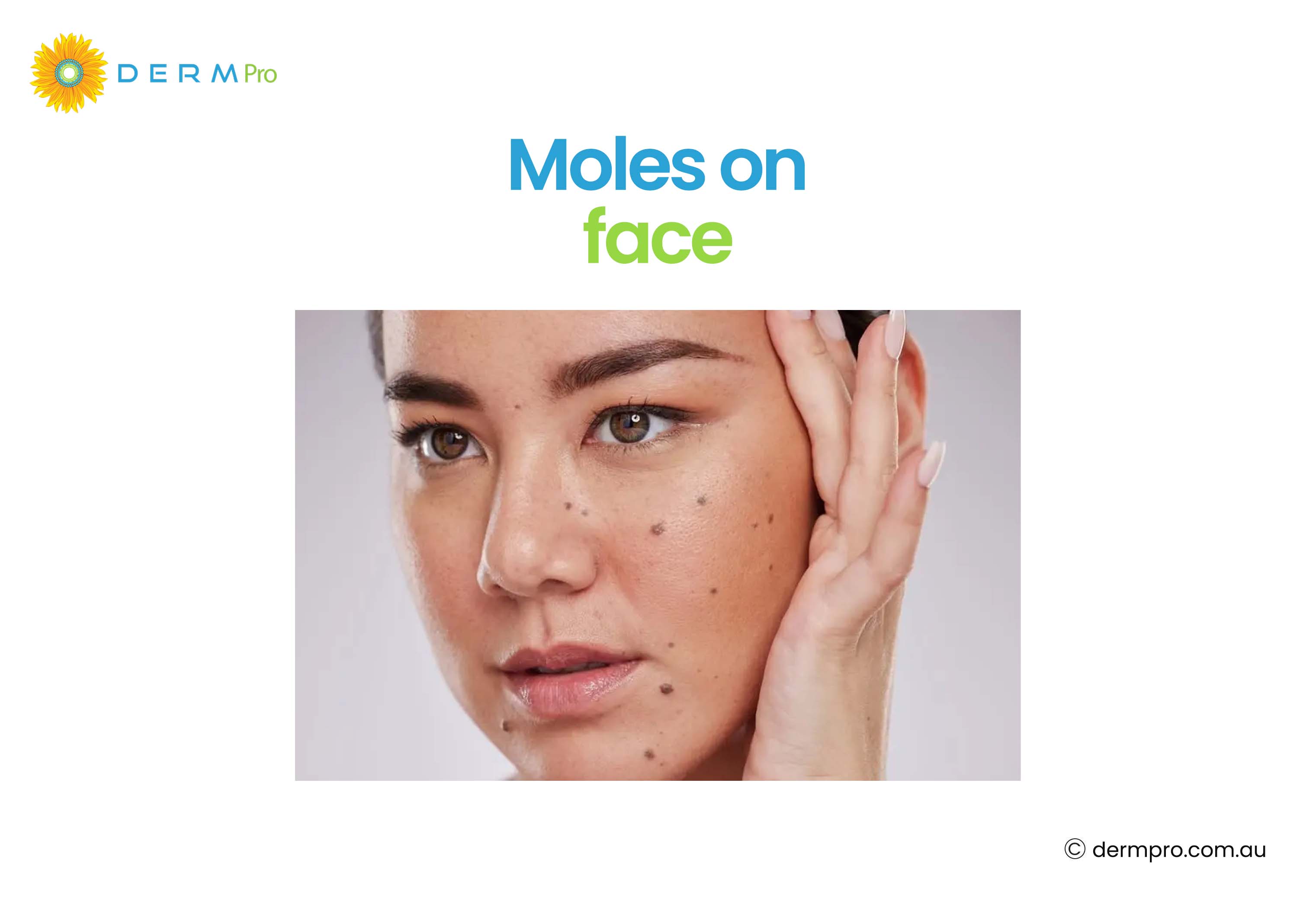
Cosmetically, many people choose to have facial moles removed. Medically, a mole on the face should be assessed for any changes in size, colour, or shape before removal to ensure it is benign.
Warning Signs of Suspicious Moles and Skin Cancer
Knowing suspicious moles and skin cancer warning signs is crucial to keeping your skin healthy. The ABCDE rule is the most widely used guide for identifying potentially concerning moles.
The ABCDE Rule helps identify warning signs of possible skin cancer or melanoma:
- A – Asymmetry: Mole one-half does not match the other
- B – Border: Mole edges are blurred, irregular and uneven
- C – Colour: Different colours or uneven colour throughout
- D – Diameter: Growing in size and can be larger than 6mm
- E – Evolving: Mole changes in colour, shape and size or symptoms over time
Not all melanomas follow the ABCDE rule. Some melanomas may appear as a new spot that looks different from other moles, sometimes called the 'ugly duckling' sign.
What Type of Cancer Do Atypical Moles Turn Into?
The type of cancer that atypical moles can turn into is melanoma. It is a form of skin cancer that begins in the melanocytes. Having dysplastic naevi does not mean a person will develop melanoma, but it does increase the risk. People with many atypical moles, a family history of melanoma, or a personal history of sunburn have a higher overall risk.
Another type of melanoma worth knowing about is acral lentiginous melanoma. This is a less common form that develops on the palms, soles of the feet, and under the nails. It can affect people of any skin tone and is sometimes harder to detect because of its location. A mole on a finger or under the nail that looks unusual should be assessed promptly.
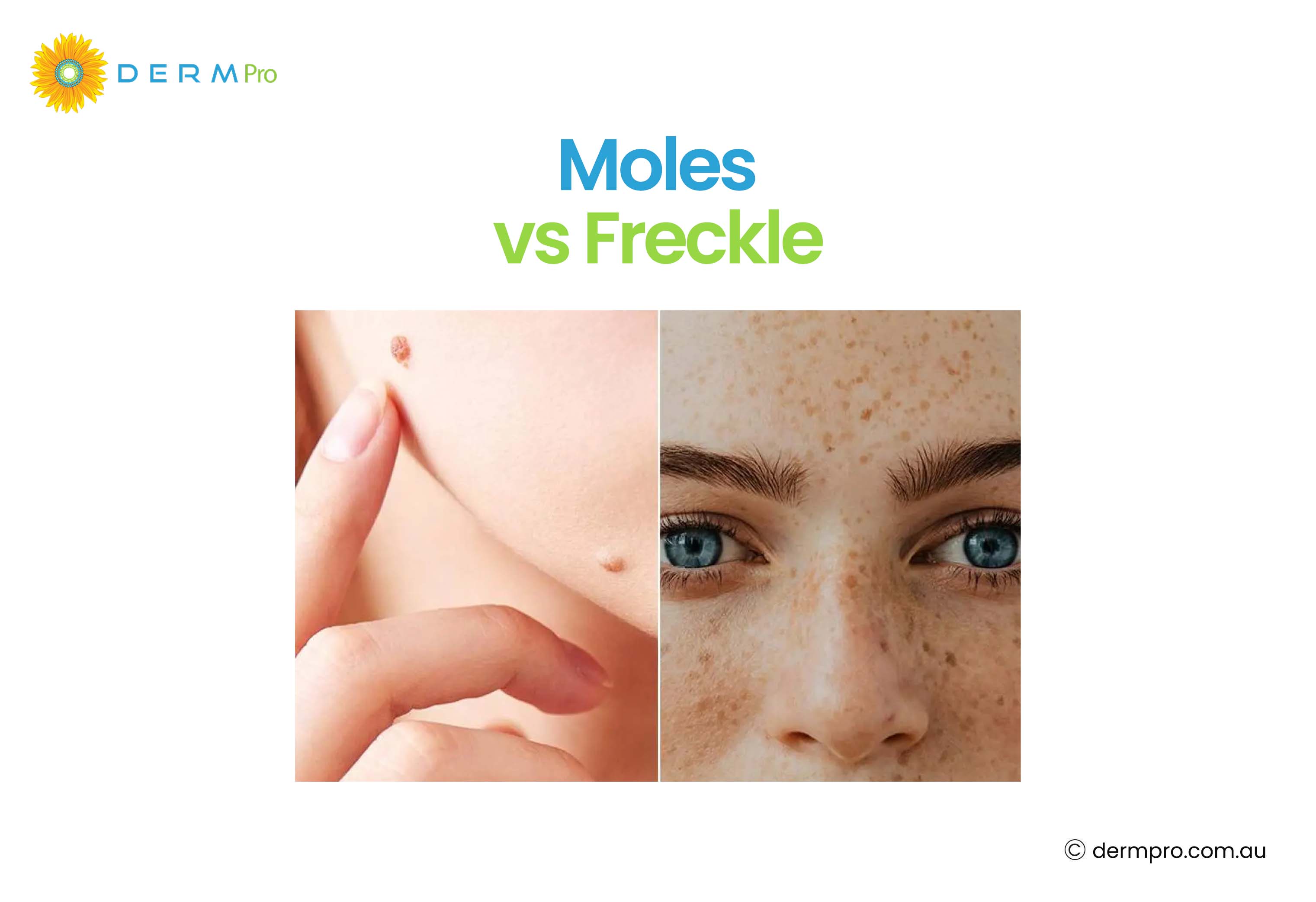
Mole vs Freckle: What Is the Difference?
The difference between moles and freckles is confusing. It is because both often look similar, especially on lighter skin. However, both moles and freckles develop in different ways and also have different features.
|
Feature |
Mole |
Freckle |
|
Cause |
Cluster of melanocytes |
Increased melanin in existing cells |
|
Appearance |
Flat, darker and raised |
Flat, small, light brown |
|
Presence at birth |
Sometimes (congenital) |
Rarely |
|
Sun impact |
Can become darker with the sun |
Fade in winter while darkening in summer |
|
Permanence |
Permanent |
Can fade with age |
|
Cancer risk |
Very low (most benign) |
It is not cancer |
Mostly, both freckles and moles are harmless. The mole vs freckle distinction is crucial to monitor for skin changes over time.
Mole vs Freckle Image
Other Skin Lesions Often Confused With Moles
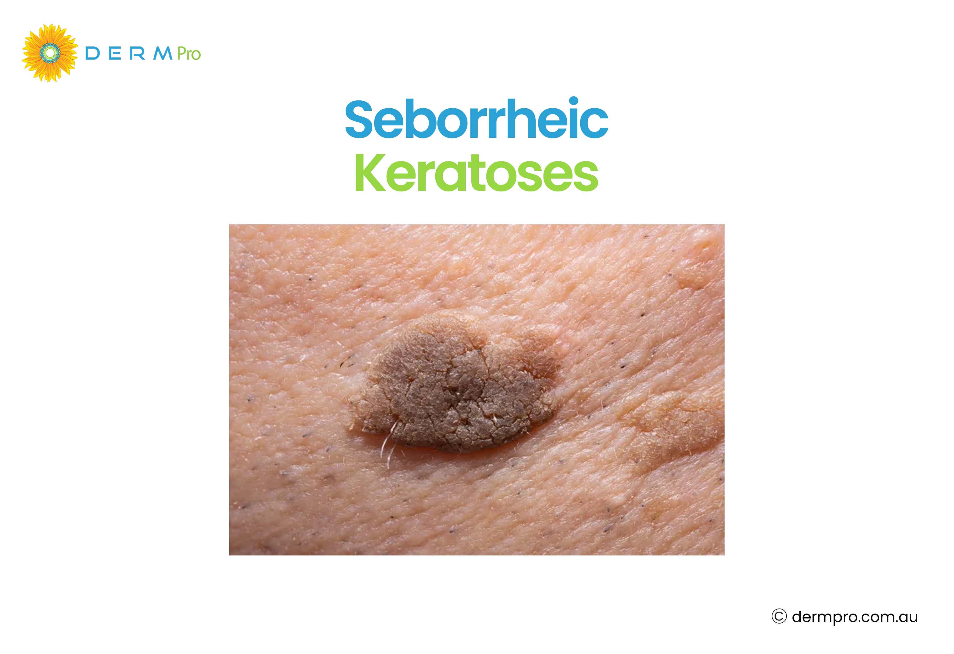
Seborrheic Keratoses
Seborrheic keratoses are non-cancerous, but they are very common. They often appear in middle age and beyond. They have a waxy and stuck-on look. However, they are brown, tan or black in colour. Many people worry about them because they can look dark and irregular.
When it comes to seborrheic keratosis vs melanoma, the key differences are texture and surface. Seborrheic keratoses have a rough and warty surface. But melanoma tends to be smoother with more variable pigmentation. However, if you are unsure, always get a lesion checked by a professional.
Seborrheic Keratoses
Lentigines
Lentigines (singular: lentigo) are flat and brown spots. They appear on sun-exposed areas of skin. They are also known as age spots or liver spots. Unlike freckles, they do not fade in winter. They are generally benign but can occasionally be confused with lentigo maligna, an early form of melanoma, which is why persistent, growing, or changing spots should be assessed. Solar lentigines become more common with age and cumulative sun exposure.
Lentigines Image

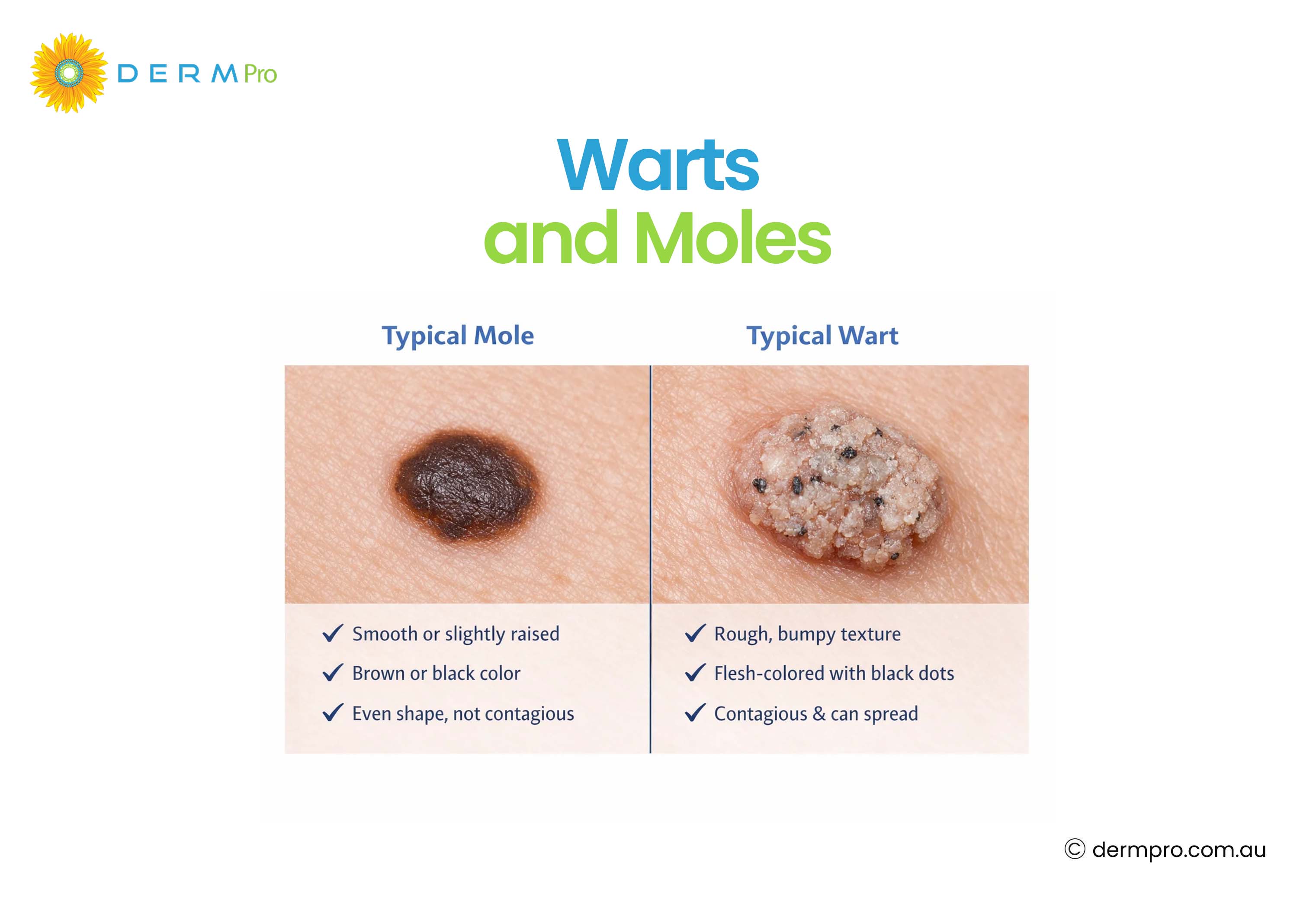
Warts and Moles
Both these terms are often confused by people. Warts are caused by the human papillomavirus (HPV) and have a rough, cauliflower-like surface. Moles are made of melanocytes and tend to be smoother. If you have a growth on the skin and are not sure whether it is a wart or a mole, a dermatologist can identify it quickly.
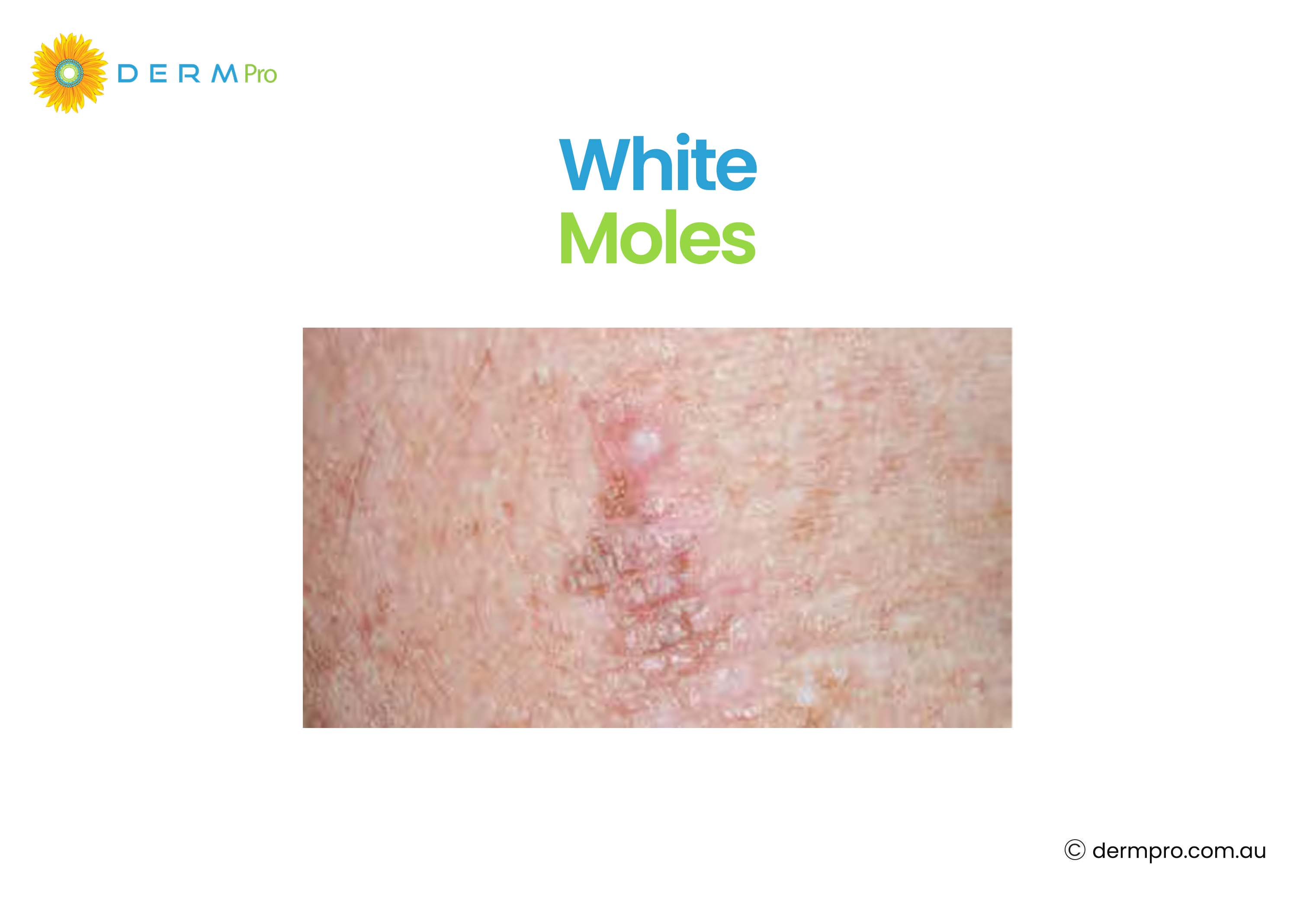
White Mole
A white mole can occur when a mole loses its pigment, a process called halo naevus. A pale ring or white area around a mole forms as the immune system targets the melanocytes. This is usually harmless, but a white mole or any new white area around a mole should be checked, as depigmentation can occasionally be associated with other conditions.
White Mole
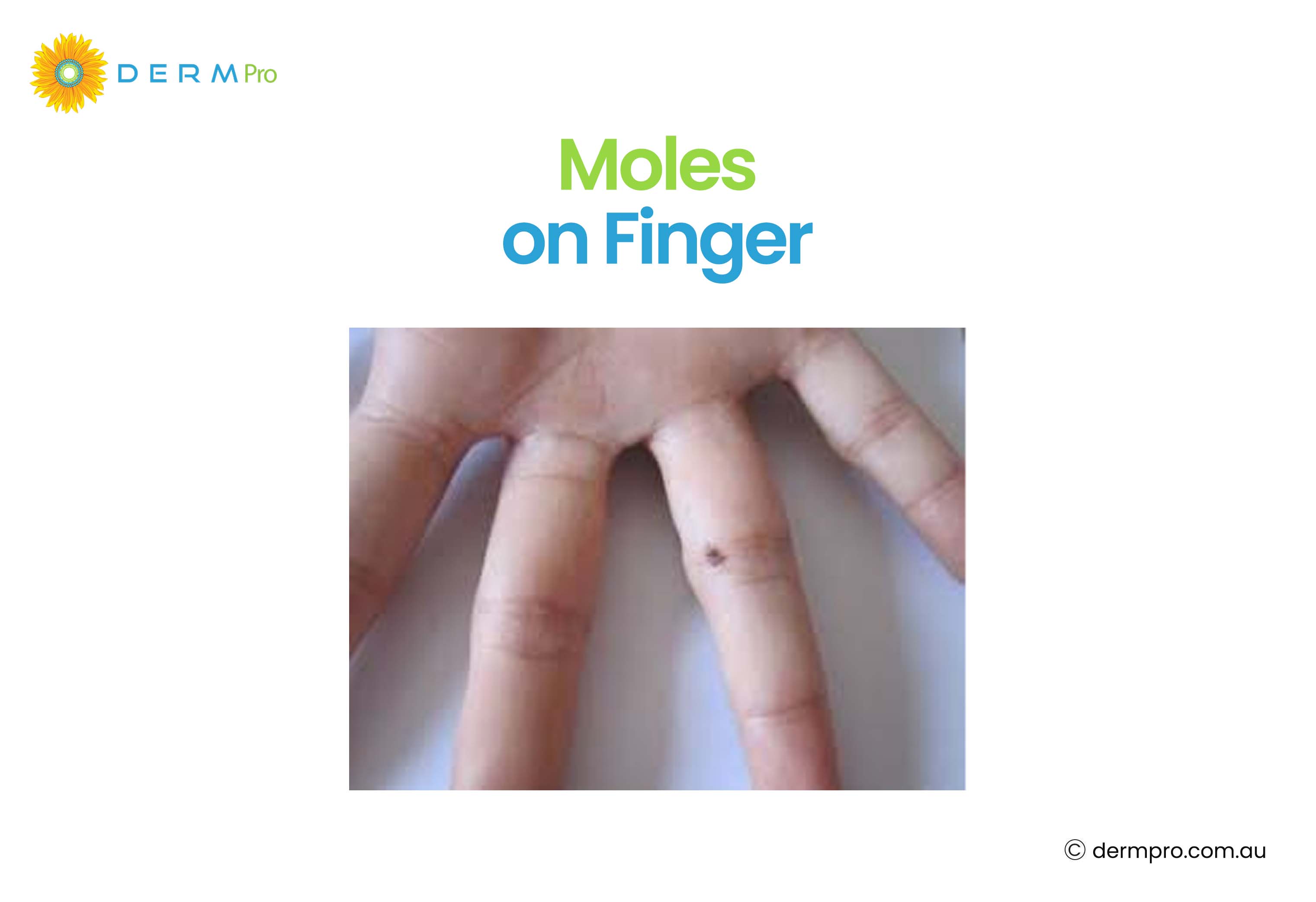
Mole on Finger
A mole on the finger is worth monitoring carefully. Moles in unusual locations, particularly on the fingers, toes, under the nails, or palms, can sometimes be associated with acral lentiginous melanoma. Any dark streak under a nail or a new mole on the hands or feet that changes should be assessed by a doctor. Most moles on the fingers are benign, but lesions in acral areas should be monitored carefully for change.
Mole on Finger
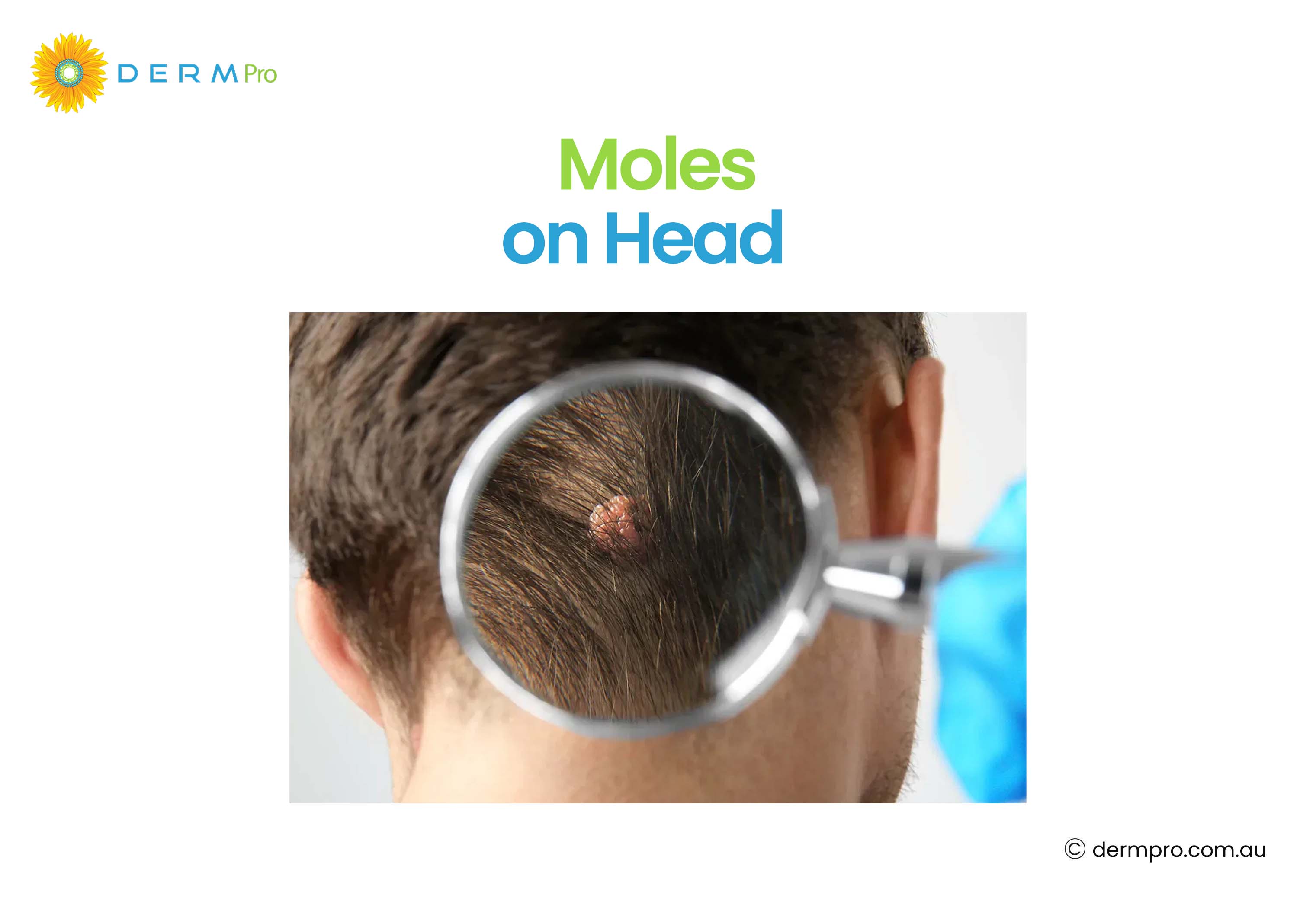
Mole on Head
A mole on the head can be hard to see without assistance. Regular scalp checks, especially for people with thinning hair or who shave their heads, are important. Partners, barbers, or hairdressers sometimes notice these first. Any mole on the scalp that bleeds, grows, or changes should be reviewed by a healthcare professional.
Mole on Head
Diagnosis and Skin Checks
When to See a Doctor?
You should see a doctor or dermatologist if a mole or skin lesion:
- Changes in colour, size, or shape
- Has an irregular or uneven border
- Bleeds or oozes without being injured
- Itches or feels tender
- Has been present since birth and is larger than 20 cm
- Appears new after the age of 40
Dermoscopy
Dermoscopy is a non-invasive technique used by dermatologists and trained GPs to examine moles more closely. A handheld device called a dermatoscope uses polarised light to reveal structures within the skin that are not visible to the naked eye. It significantly improves the accuracy of identifying suspicious moles.
Skin Imaging
Total body photography and skin imaging allow a doctor to document and track all moles over time. This makes it easier to detect changes at follow-up appointments. Digital dermoscopy platforms and AI-assisted tools support this process.
Why Regular Monitoring Matters?
Skin cancer is one of the most common cancers in Australia. The good news is that most skin cancers, including melanoma, have much better outcomes when detected early. Regular self-checks and professional skin checks are the best way to catch changes before they progress.
Treatment Options
There are multiple treatment options depending on the type of skin cancer. The most common are:
Monitoring Only
Most benign moles require no treatment at all. A doctor may recommend a watch-and-wait approach, taking photographs to document the mole and reviewing it at regular intervals.
Surgical Removal
Surgical removal is recommended for suspicious moles that have changed or are causing physical discomfort. This involves a minor procedure under local anaesthetic where the mole is excised and sent for biopsy. Removal is usually straightforward and leaves a small scar.
Cryotherapy for Seborrheic Keratoses
Cryotherapy for seborrheic keratoses uses liquid nitrogen to freeze and remove the growths. It is a quick, effective, and commonly used treatment. Multiple lesions can be treated in one session. Seborrheic keratosis treatment also includes curettage (scraping) and laser therapy, depending on the size and location of the lesions.
How DermPro Supports Early Skin Monitoring?
Understanding your skin is the first step in protecting it. DermPro is an Australian AI skin health platform designed to help people track and monitor suspicious skin lesions over time.
DermPro is not a diagnostic tool and does not replace the assessment of a qualified healthcare professional. Rather, it supports earlier skin awareness by encouraging people to pay attention to their skin regularly and giving them a way to document changes. This can lead to more informed conversations with a GP or dermatologist.
If you want to start tracking your moles and taking your skin health more seriously, DermPro offers a simple way to begin.
Conclusion
Moles and skin lesions come in many forms. Understanding the types of moles on skin, including benign moles, congenital naevi, dysplastic naevi, and atypical moles, helps you know what to look for and when to seek advice.
Most moles on skin are completely harmless and never need treatment. But skin is always changing, and some changes are worth taking seriously. The ABCDE rule, regular self-checks, and professional skin assessments are all part of staying on top of your skin health.
If you notice any mole that is new, changing, or just not quite right, do not wait. Early assessment is always the best approach. And if you want help staying consistent with tracking your skin between appointments, tools like DermPro are a practical place to start.
Frequently Asked Questions
How many different types of moles are there?
There are many types of moles, including benign moles, congenital naevi, acquired melanocytic naevi, compound naevi, intradermal naevi, junctional melanocytic naevi, blue naevi, Spitz naevi, and dysplastic (atypical) naevi with varying grades of atypia.
What is the difference between a mole and a freckle?
Moles are clusters of melanocytes that can be raised or flat and are permanent. Freckles are flat and lighter spots caused by increased melanin production that often fade in winter. Freckles are not associated with cancer risk.
What type of cancer does an atypical mole turn into?
Atypical moles (dysplastic naevi) can, in some cases, develop into melanoma, a type of skin cancer. However, most atypical moles never become cancerous. People with multiple atypical moles have a higher overall melanoma risk and benefit from regular skin checks.
What is seborrheic keratosis, and is it dangerous?
Seborrheic keratosis is a common and non-cancerous skin growth with a waxy, stuck-on appearance. It is not dangerous, but it can resemble melanoma. Any lesion that is growing, changing, or unclear should be assessed by a healthcare professional.
When should I see a doctor about a mole?
See a doctor if a mole changes in colour, size, or shape, develops an irregular border, bleeds, itches, or is new after age 40. The ABCDE rule is a helpful guide for identifying suspicious moles.
How does DermPro help with skin monitoring?
DermPro is an Australian AI skin health platform that allows users to photograph and track skin lesions over time. It supports early awareness and helps users have more informed conversations with their doctor. It is not a diagnostic tool.
What is acral lentiginous melanoma?
Acral lentiginous melanoma is a less common form of melanoma that develops on the palms, soles of feet, or under the nails. It can affect people of any skin tone and often presents as a dark streak under a nail or a growing spot on the foot or hand.
This blog is for informational purposes only and does not replace professional medical advice, diagnosis, or treatment. Always consult a qualified healthcare professional for proper evaluation of any skin changes or concerns.
Was this article helpful?
Your feedback helps us create better content for everyone.